Service
Advanced Medical Imaging
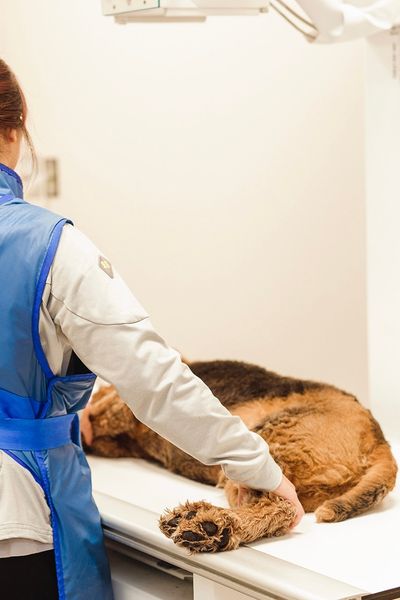
Our hospital offers medical imaging services, which are an important tool for diagnosing many diseases. Our facility has a variety of equipment to meet different needs: digital radiographic imaging (x-ray), ultrasound imaging (ultrasound) and coming soon, CT-Scan (computed tomography, x-ray) imaging.

DIGITAL RADIOGRAPHIC IMAGING (X-RAY)
Veterinary medical imaging is an important tool in the diagnosis of many of our pets’ diseases:
- Upper and lower limb radiographs for bone, intervertebral disc, or joint lesions.
- Neck radiographs for upper respiratory tract diseases (e.g. tracheal, laryngeal and oesophageal injuries).
- Thoracic (3 views) radiographs for lung, heart, and blood vessel lesions.
- Abdominal radiographs (3 views) for the stomach, intestine, liver, spleen, kidneys, bladder and tumours.
We have a digital radiography system that allows us to produce very detailed radiographs quickly. Digital technology allows the image to be computer enhanced for optimal diagnostic benefit. In addition, the radiographs can be saved on CD-ROM or USB stick.
Referral to a radiographic interpretation service by board-certified ACVR radiologists is included in the price of all emergency radiographic studies. An official report is provided for the medical record.
ECHOGRAPHY IMAGING (ULTRASOUND)
Our Ottawa hospital also has veterinary medical imaging, including an ultrasound unit allowing diagnostic imaging above and beyond abdominal and thoracic radiographs. This is particularly useful for quickly assessing the thoracic and abdominal cavities and their organs for fluid, masses, or other abnormalities.
Ultrasonography is most frequently used to investigate the abdomen but can also be used to evaluate other anatomic regions, such as the thorax. During ultrasound examinations, fine-needle aspirates can also be performed to help obtain a diagnosis on masses and lesions. Ultrasound also allows precise sampling of sterile urine from the bladder to submit for bacterial culture in case of potential urinary infection.
Safe light sedation (or heavier but reversible sedation) is usually required to allow proper ultrasound assessment of lesions and organs and to allow safe sampling from needle aspirations.
CT-SCAN (COMPUTED TOMOGRAPHY, X-RAY) IMAGING
CT scanning is done under heavy reversible sedation or general anesthesia and is used to diagnose or follow the progression of many diseases, such as complex fractures, vertebral disc herniation, nasal cavity diseases, chronic ear problems, pre-surgical evaluation of masses, and whole-body scanning for the staging of cancer patients.

